File:Bundleofhis.png
Bundleofhis.png (400 × 483 pixel, dimensione del file: 69 KB, tipo MIME: image/png)
| Questo file e la sua pagina di descrizione (discussione · modifica) si trovano su Wikimedia Commons (?) |
Dettagli
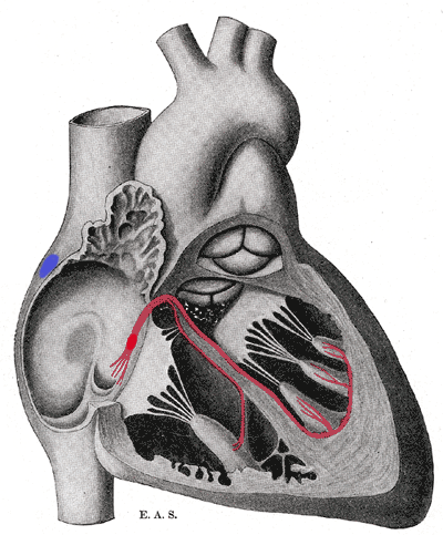
Schematic representation of the atrioventricular bundle of His. The bundle, represented in red, originates near the orifice of the coronary sinus, undergoes slight enlargement to form the AV node. The AV node tapers down into the bundle of HIS, which passes into the ventricular septum and divides into two bundle branches, the left and right bundles. Sometimes the 'left and right bundles of His' are called Purkyne or Purkinge fibres. The ultimate distribution cannot be completely shown in this diagram.
This image is misleading. Although it correctly places the SA and AV nodes in the right atrium, it appears as though there are only two papillary muscles in the right ventricle and three in the left ventricle. The opposite is actually true. The papillary muscles attach to chordae tendinae which then attach to the leaflets of the AV valves, preventing prolapse. The left AV valve is the mitral or bicuspid and only has two leaflets and therefore two papillary muscles. The right AV valve is the tricuspid and should have three papillary muscles corresponding to the three leaflets of the valve.
Licenza
Questo file è nel pubblico dominio negli Stati Uniti. Questo si applica alle opere statunitensi il cui copyright è scaduto, spesso perché la sua prima pubblicazione è avvenuta prima del 1º gennaio 1929. Vedi questa pagina per ulteriori spiegazioni.
|
 | |
 |
Questa immagine potrebbe non essere nel pubblico dominio al di fuori degli Stati Uniti, in particolare nei Paesi e nelle aree in cui non viene applicata la regola della durata più breve per le opere statunitensi, come in Canada, Cina (non a Hong Kong, Macao o Taiwan), Germania, Messico e Svizzera. Il creatore e l’anno di pubblicazione sono informazioni essenziali e devono essere fornite. Vedi Wikipedia:Public domain e Wikipedia:Copyright per maggiori dettagli.
|
Cronologia del file
Fare clic su un gruppo data/ora per vedere il file come si presentava nel momento indicato.
| Data/Ora | Miniatura | Dimensioni | Utente | Commento | |
|---|---|---|---|---|---|
| attuale | 19:40, 20 set 2006 | | 400 × 483 (69 KB) | Kauczuk | Bundle of His, from Gray's Anatomy 1918 |
Pagine che usano questo file
La seguente pagina usa questo file:
Utilizzo globale del file
Anche i seguenti wiki usano questo file:
- Usato nelle seguenti pagine di ar.wikipedia.org:
- Usato nelle seguenti pagine di az.wikipedia.org:
- Usato nelle seguenti pagine di bn.wikibooks.org:
- Usato nelle seguenti pagine di bs.wikipedia.org:
- Usato nelle seguenti pagine di ca.wikipedia.org:
- Usato nelle seguenti pagine di cs.wikipedia.org:
- Usato nelle seguenti pagine di de.wikipedia.org:
- Usato nelle seguenti pagine di de.wikibooks.org:
- Usato nelle seguenti pagine di el.wikipedia.org:
- Usato nelle seguenti pagine di en.wikipedia.org:
- Usato nelle seguenti pagine di en.wikibooks.org:
- Usato nelle seguenti pagine di es.wikipedia.org:
- Usato nelle seguenti pagine di es.wikibooks.org:
- Usato nelle seguenti pagine di eu.wikipedia.org:
- Usato nelle seguenti pagine di fr.wikipedia.org:
- Usato nelle seguenti pagine di hi.wikipedia.org:
- Usato nelle seguenti pagine di ja.wikipedia.org:
- Usato nelle seguenti pagine di ja.wikibooks.org:
- Usato nelle seguenti pagine di ko.wikipedia.org:
- Usato nelle seguenti pagine di lv.wikipedia.org:
- Usato nelle seguenti pagine di nl.wikipedia.org:
- Usato nelle seguenti pagine di pl.wikipedia.org:
- Usato nelle seguenti pagine di pt.wikipedia.org:
- Usato nelle seguenti pagine di sr.wikipedia.org:
- Usato nelle seguenti pagine di www.wikidata.org: