File:P450cycle.svg

File originale (file in formato SVG, dimensioni nominali 9 240 × 6 968 pixel, dimensione del file: 38 KB)
| Questo file e la sua pagina di descrizione (discussione · modifica) si trovano su Wikimedia Commons (?) |
Dettagli
| Descrizione |
English: ==The P450 catalytic cycle==
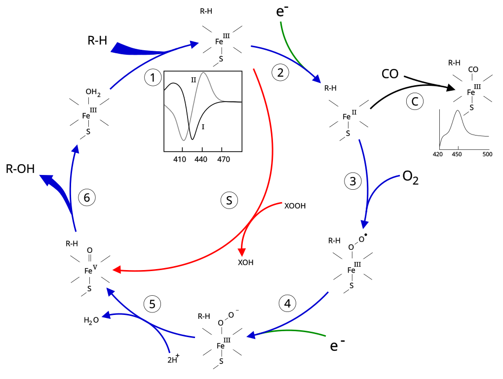
1: The substrate binds to the active site of the enzyme, in close proximity to the heme group, on the side opposite to the peptide chain. The bound substrate induces a change in the conformation of the active site, displacing a water molecule from the distal axial coordination position of the heme iron[1] changing the state of the heme iron from low-spin to high-spin[2]. This gives rise to a change in the spectral properties of the enzyme, with an increase in absorbance at 390~nm and a decrease at 420~nm. This can be measured by difference spectrometry and is referred to as the "type~I" difference spectrum (see inset graph in figure). Some substrates cause an opposite change in spectral properties, a "reverse type~I" spectrum, by processes that are as yet unclear. Inhibitors and certain substrates that bind directly to the heme iron give rise to the type~II difference spectrum, with a maximum at 430~nm and a minimum at 390~nm (see inset graph in figure). If no reducing equivalents are available, this complex remains stable, allowing the degree of binding to be determined from absorbance measurements in vitro[3] 2: The change in the electronic state of the active site favours the transfer of an electron from NAD(P)H[4]. This takes place via the electron transfer chain, as described above, reducing the ferric heme iron to the ferrous state. 3: Molecular oxygen binds covalently to the distal axial coordination position of the heme iron. The cysteine ligand is a better electron donor than histidine, with the oxygen consequently being activated to a greater extent than in other heme proteins. However, this sometimes allows the bond to dissociate, the so-called "decoupling reaction", releasing a reactive superoxide radical, interrupting the catalytic cycle[1]. 4: A second electron is transferred via the electron-transport system, reducing the dioxygen adduct to a negatively charged peroxo group. This is a short-lived intermediate state. 5: The peroxo group formed in step 4 is rapidly protonated twice by local transfer from surrounding amino-acid side chains, releasing one mole of water, and forming a highly reactive iron(V)-oxo species[1]. 6: Depending on the substrate and enzyme involved, P450 enzymes can catalyse any of a wide variety of reactions. A hypothetical hydroxylation is shown in this illustration. After the product has been released from the active site, the enzyme returns to its original state, with a water molecule returning to occupy the distal coordination position of the iron nucleus. S An alternative route for mono-oxygenation is via the "peroxide shunt": interaction with single-oxygen donors such as peroxides and hypochlorites can lead directly to the formation of the iron-oxo intermediate, allowing the catalytic cycle to be completed without going through steps 3, 4 and 5[3]. A hypothetical peroxide "XOOH" is shown in the diagram. C: If carbon monoxide (CO) binds to reduced P450, the catalytic cycle is interrupted. This reaction yields the classic CO difference spectrum with a maximum at 450 nm.
|
| Data | |
| Fonte | M.Sc. Thesis, David Richfield (User:Slashme) |
| Autore |
Slashme di Wikipedia in inglese When using this image in external works, it may be cited as follows:
|
Licenza
| Io, detentore del copyright su quest'opera, la rilascio nel pubblico dominio. Questa norma si applica in tutto il mondo. In alcuni paesi questo potrebbe non essere legalmente possibile. In tal caso: Garantisco a chiunque il diritto di utilizzare quest'opera per qualsiasi scopo, senza alcuna condizione, a meno che tali condizioni siano richieste dalla legge. |
Cronologia del file
Fare clic su un gruppo data/ora per vedere il file come si presentava nel momento indicato.
| Data/Ora | Miniatura | Dimensioni | Utente | Commento | |
|---|---|---|---|---|---|
| attuale | 17:07, 22 apr 2012 | | 9 240 × 6 968 (38 KB) | Slashme | {{Information |Description ={{en|1====The P450 catalytic cycle== 1: The substrate binds to the active site of the enzyme, in close proximity to the heme group, on the side opposite to the peptide chain. The bound substrate induces a change in the ... |
| 16:49, 22 apr 2012 | Nessuna miniatura | 9 240 × 6 968 (38 KB) | Slashme | Corrected peroxide shunt arrow. | |
| 12:34, 5 lug 2008 |  | 9 240 × 6 968 (35 KB) | Slashme | {{Information |Description={{en|1===The P450 catalytic cycle== 1: The substrate binds to the active site of the enzyme, in close proximity to the heme group, on the side opposite to the peptide chain. The bound substrate induces a change in the conforma |
Pagine che usano questo file
Le seguenti 2 pagine usano questo file:
Utilizzo globale del file
Anche i seguenti wiki usano questo file:
- Usato nelle seguenti pagine di bs.wikipedia.org:
- Usato nelle seguenti pagine di ca.wikipedia.org:
- Usato nelle seguenti pagine di de.wikipedia.org:
- Usato nelle seguenti pagine di el.wikipedia.org:
- Usato nelle seguenti pagine di en.wikipedia.org:
- Usato nelle seguenti pagine di en.wikibooks.org:
- Usato nelle seguenti pagine di en.wikiversity.org:
- Usato nelle seguenti pagine di fa.wikipedia.org:
- Usato nelle seguenti pagine di gl.wikipedia.org:
- Usato nelle seguenti pagine di he.wikipedia.org:
- Usato nelle seguenti pagine di la.wikipedia.org:
- Usato nelle seguenti pagine di ru.wikipedia.org:
- Usato nelle seguenti pagine di zh.wikipedia.org: